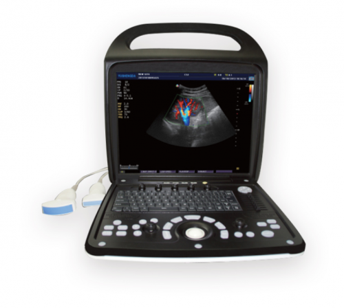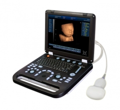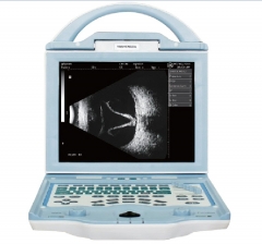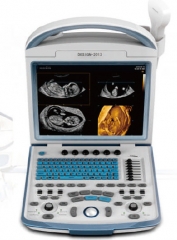

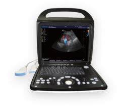
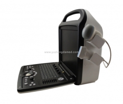

YSD900B is premium full digital ultrasound diagnostic instrument that provides a comprehensive diagnostic solution to our valuable users in all types of clinical environments. 15 inches LED monitor,ergonomic rotatable design keyboard and four probe connectors make the users easy to operate.
The wide range of applications include Abdomen, OB/GYN, urology, andrology, cardiology, etc.
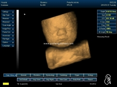
Meanwhile this machine can be updated to 4D software.
Main Feature
1.Easy to carry only 5KG
2.Powerful software packages almost includes most of part
B, B|B, 4b, B|M, M, PW, CW, CFM, PDI, DPDI, Duplex, 3D&4D imaging
3.Human-kind design,Operation easily.
4.15'' High resolution LED display.
Imaging Modes:
B, B|B, 4B, B|M, M
Color Doppler (CFM)
Power Doppler (PDI)
Directional Power Doppler (DPDI)
Pulsed Wave Doppler (PWD)
B+PWD (Duplex)
B+CFM/PDI/DPDI+PWD (Triplex)
High Pulse Repetition Frequency (HPRF)
Tissue Harmonic Imaging (THI) Scanning Method:
Electronic linear, electronic convex, electronic micro-convex
Detail Feature Sheet
Weighing less than 5 kgs, comfortably pick and go. |
Built-in battery, working time more than 2 hours, extending point of care to sites where power sources are unavailable. |
Not only a professional ultrasound system, could also be a laptop computer upon request. |
15 inch LED display, as large as 175 degree viewing angle. |
8-segment TGC, precise adjustment of near/far gain. |
2 usb ports, connectivity of usb flash and laser printer |
Discom 3.0 port, compatibility with archives, PACS or serves |
Projector port, a must for lecturing or training |
Probe variety: convex, micro-convex, endo-cavity/rectal, linear, volume |
Specialty: general, OB/GYN, vascular, cardiology, urinary, small organs, etc. |
Display mode; B, 2B, 4B, B/M, M |
Imaging mode: 2D, free-hand 3D, 4D |
Image/video format: AVI, JPG, BMP, PNG, TIF, DICOM |
Image processing
Brightness, contrast
Advanced gamma control
Scan direction, rotation
Black/white, Left/right revers e
Up/down, negative / positive control
8 segments TGC Control
High Pulse Repetition Frequency (HPRF)
Tissue Harmonic Imaging (THI)
PSHI TM broadband multi-frequency harmonic image
Speckle-reduction
Noise rejection function
Power adjustable
iBeam TM intelligent space image technology
iZoom TM undistorted full screen image
iClear
iTouch
Edge enhancement
Image conversion
Doppler Sound output volume adjustable
Color threshold control
Wall filter adjustable
Base line adjustable
Sampling frame adjustable
Spectrum sampling volume adjustable
Spectrum sampling volume angle adjustable
Stereo sound: Volume control
PRF adjustable

 Functions:
Cine loop: more than 512 frame cine loop memory
Storage media: 500G massive image-storage capacity
Zoom: Pan zoom
USB ports: 2
Scanning Method
Electronic linear, electronic convex, electronic micro-convex, scanning depth: 2-30cm
Doppler ultrasound scanner
· PRF variable: 0.5-10 kHz
· Wall filter settings: 3 steps (5%, %10%, 15% PRF)
· Angle steering for linear transducers: ±10°
· Real-time spatial filter: 4 values
· CFM palette>10 maps
· PDI palette>10 maps
· B/Color priority control
· Color threshold control
· CFM baseline control
· Doppler frequency selection
· Color frame averaging
· Transparent Color Mapping (TCM)
Pulsed wave doppler
· PRF variable: 1-10 kHz
· Wall filter settings: 16 steps (2.5%-20% PRF)
· Angle steering for linear transducers: ±10°
· Real-time trace line with automatic calculation of spectrum parameters
· Stereo sound: volume control
· PWD palette>10 maps
· Doppler frequency selection
Processing
· High Line Density scan mode for better resolution
· 8 sliders TGC Control
· Dynamic range>120 dB
· Overall gain control
· M - mode sweep speed control
· Acoustic power control
· Variable frame averaging
· Brightness, contrast
· Advanced gamma control
· Scan direction, rotation, up-down controls
· Negative / positive control
· Echo enhancement control
· Noise rejection function
· Speckle reduction
Specifications:
Display | Full digital 15 inch LED display, 4D ultrasound |
Platform | Pc-based |
Probes | 3.5MHz R60/R50 convex probe; multi-frequency from 2.0MHz to 5.5MHz 7.5MHz L40 linear probe; multi-frequency from 5.0MHz to 14.0MHz 6.5MHz R10/R13 transvaginal probe; multi-frequency from 5.0MHz to 9.0MHz 3.5MHz R20 cardiac probe; multi-frequency from 2.0MHz to 5.5MHz |
Beam forming | DBF, RDA, DRA, DRF |
DFS | Dynamic frequency scanning from 2.0 to 12.0MHz, 4 multi-frequency scanning |
THI | Yes |
Display mode | B, 2B, 4B, B/M, M |
Dynamic range | ≥100dB, 4 steps of switching functions |
Image process technologies | controllable frame correlation, Gamma correction, edge enhancement, image smoothing, image denoisiong, automatical gain adjustment, up/down, left/right and black/white conversation. |
Image magnification | stepless magnification, dynamic real-time PIP local zoom functions |
Cine loop | 1024 frame auto/manual cine loop; multi screens cine loop (4B, 9B); auto/manual cine loop under B/M and M mode. |
Image management system | the functions of pigeonholing, browsing, comparing, saving, printing and transferring images; as many as hundreds of thousands of images and thousands of cine loop could be saved; saved images could be operated by full-screen browse under slide mode. |
Measurement and calculation | measure perimeter and area by distance or ellipse method; measure perimeter and area by track method; measure body surface area and volume by ellipse method. 4 measure sticks; rate measure; linear stenosis ratio, area stenosis ratio, angle measure. All calculations are automatic. |
Assist tools | puncture guide, histogram, sectional drawing. |
Menu manage interface | real time online support and navigation clew system, image fore-set and one-key optimization functions. |
Body marks | Multi-tens |
Patient cases database system | All the data could be saved, searched and managed. Multiple kinds of OB. measurement repors, fetus physiological grades and repors and fetus growth curve. |
Presetting Formulas | Presetting system for diagnosis and measurement formulas. Different formulas could be set according to different races. |
Auto-measure software of OB., Gyn., small organs, cardiac, urology and others | OB.: BPD, CRL, GS, HA, AC, HC, FL, APAD, TAD, FTA, HUMERUS, OFD, THD, TIBIA, ULNA, AFI, LIMP, BBT, FBP Gyn.: uterus diameter, intima thickness, ovary colume, regnant ovarian follicle, length of cervix long-diameter, uterine. Small organs: thyroid gland, hip joint. Cardiac: AOD, LAD, IVSTd, LVIDd, AA, LAD/AOD, LVPWd, LVIDs, EF, EF SLP, CA/CE, MVCF, CO, CI, LVMWI, AVSV, FS, ACV, ET, SV, SI, LVMW, QMV. Urology: remained urine sample, prostate,PSAD. Patient cases database systems. All the data could be saved, searched and managed. Multiple kinds of OB. measurement repors, fetus physiological grades and repors and fetus growth curve. |
3D software | Yes |
4D software | Yes, optional |
Relative extended ports | VGA, S-Video, TV video port USB2.0 port, 2G saving card RJ-45 network port Multiple kinds of saving modes are all supported, containing soft disk, hard disk, flash disk, CF card, SD card and others. Compatible of jet printer, laser printer, video printer and video recorder |
Expansion interfaces
VGA, TV Interface
USB2.0 Interface
RJ-45 Network interface
Support DeskJet printer, LaserJet printer, video printer
Transducers
2-5 MHz Electronic convex array transducer
5-14 MHz Electronic linear array transducer
2-5 MHz Electronic micro-convex transducer
5-10 MHz Electronic transvaginal array transducer
Standard Configuration:
Main Unit,
15 inch LED monitor,
frame arm,
3.5 MHz convex probe,
7.5MHz linear probe,
2 probe connectors,
2 USB port,
Hard Disc (500G)
Options:
6.5 MHz transvaginal probe,
3.5MHz microconvex probe,
3.0 MHz phased array probe,
4D volume probe,
laser printer,
video printer,
DICOM 3.0, DVD-RW
Probe information:
Probes | |
| 3.5Mhz abdominal probe | 2.0, 3.0, 3.5, 4.0, 5.5Mhz |
| 6.5Mhz transvaginal probe | 5.0, 6.0, 6.5, 7.5, 9.0Mhz |
| 7.5Mhz linear probe | 6.0, 6.5, 7.5, 10.0, 12.0 14.0Mhz |
| 3.5MHz micro-convex probe | 2.0, 2.5, 3.5, 4.5, 5.5Mhz |
| 7.5Mhz rectum probe | 6.0, 6.5, 7.5, 10.0, 12.0Mhz |
| 3.0Mhz phased array probe | 2.0, 3.0, 3.5, 4.0, 5.5Mhz |
| 4D volume probe | 2.0, 3.0, 3.5, 4.0, 5.5Mhz |